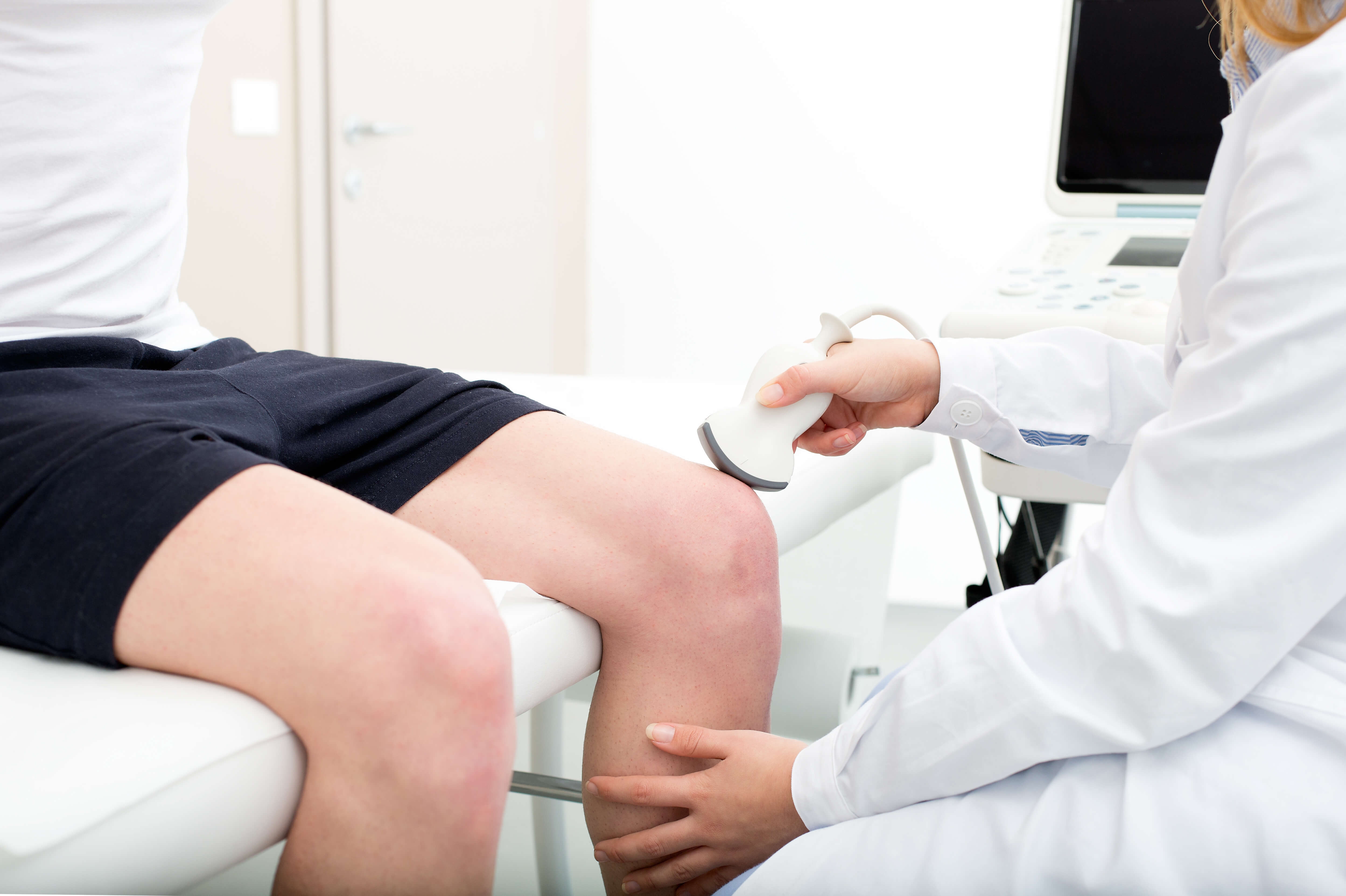
The strength of the biceps brachii muscle can be assessed manually by performing a manual muscle test. This involves applying resistance to specific movements of the arm while the individual contracts their biceps muscle. The examiner can then grade the strength of the muscle based on the individual's ability to resist the applied force.
To evaluate the function of the biceps brachii muscle, specific movements such as elbow flexion and supination of the forearm should be performed. These movements primarily engage the biceps muscle and allow the examiner to assess its strength and integrity.
I usually say, “I’ve never gotten to see Game of Thrones,” to highlight how busy my life is. For the past nineteen years, I’ve worked full-time in Emergency Services, doing shift work. Additionally, I’ve owned and operated Access Health and Fitness in Dublin with employees for twelve years. I’m also married with three level-headed, non-stress […] The post Defeating Limited Training Time: A Modified Built Strong Program appeared first on StrongFirst.
Posted by on 2024-06-20
The SFG I is a challenging certification, but if I can do it, just about anyone can. I was never the most naturally gifted athlete. But with an intelligent and consistent approach to training, I was able to turn up at the event very well prepared for what was to come. My preparation for the […] The post A+A Light—How Snatching Half Your Bodyweight Can Prepare You for the StrongFirst Kettlebell Snatch Test appeared first on StrongFirst.
Posted by on 2024-06-06
Years ago, after a Plan Strong™ seminar, I had a chance to ask Pavel a question. Me: “What do you think the biggest issue is in the fitness industry?” Pavel: “I don’t have enough time to answer that question, but I will tell you that there is far too much entertainment.” I understood exactly what […] The post Strength in Numbers: Can You Really Get Strong in a Group Fitness Class? appeared first on StrongFirst.
Posted by on 2024-05-23
Most athletes know kettlebells have many benefits, especially the ballistic skills (e.g., the swing, snatch, and clean) which are almost impossible to do with any other tool. However, what may be less known is that kettlebells give us the opportunity to improve grinds as well as develop strength in the process. One of those grind […] The post A Pressing Matter appeared first on StrongFirst.
Posted by on 2024-05-09
Common signs of weakness or injury in the biceps brachii muscle during manual assessment may include decreased strength during elbow flexion or supination, pain with resisted movements, tenderness over the muscle belly, or the presence of swelling or bruising in the area.
During manual assessment, the examiner can differentiate between a strain and a tear in the biceps brachii muscle by assessing the severity of pain, weakness, and loss of function. A strain typically involves mild to moderate pain and limited strength, while a tear may result in severe pain, significant weakness, and a visible deformity in the muscle.
Key anatomical landmarks that should be palpated during manual assessment of the biceps brachii muscle include the bicipital groove, where the tendon of the long head of the biceps inserts into the shoulder joint, and the muscle belly of the biceps in the anterior aspect of the upper arm.
Special Considerations in Manual Muscle Testing for Different Muscle Groups
A positive manual assessment result for the biceps brachii muscle may indicate normal strength and function, or it could suggest weakness, injury, or pathology within the muscle. Further diagnostic testing, such as imaging studies or referral to a specialist, may be warranted to determine the underlying cause of the positive assessment findings.
Manual assessment of the biceps brachii muscle can be integrated into a comprehensive musculoskeletal evaluation by incorporating it into a systematic assessment of the upper extremities. By evaluating the strength, range of motion, and integrity of the biceps muscle, the examiner can gather valuable information about the individual's overall musculoskeletal health and identify any potential issues that may require further evaluation or treatment.
Patient positioning is crucial in gluteus medius testing as it allows for accurate assessment of muscle strength and function. Proper positioning ensures that the muscle is isolated and can be tested effectively. This is important in diagnosing conditions such as gluteus medius weakness or dysfunction. Additionally, correct positioning helps in preventing compensatory movements or recruitment of other muscles during testing, which can lead to inaccurate results. By positioning the patient correctly, healthcare providers can obtain reliable data to inform treatment plans and monitor progress. Overall, patient positioning plays a significant role in the accuracy and effectiveness of gluteus medius testing.
Muscle weakness can be detected during manual muscle testing by assessing the individual's ability to generate force against resistance. The examiner applies pressure to specific muscle groups while the individual performs a specific movement, such as flexing or extending a joint. Weakness may be indicated by the individual's inability to maintain the desired position or movement against resistance. Other signs of muscle weakness during manual muscle testing include decreased muscle tone, reduced muscle bulk, and limited range of motion. Additionally, the presence of muscle atrophy, fasciculations, or tremors may also suggest underlying weakness. Overall, manual muscle testing is a valuable tool for evaluating muscle strength and identifying areas of weakness in individuals.
To test the gastrocnemius muscle in patients experiencing calf pain, a healthcare provider may perform a series of physical examinations. This may include assessing the range of motion of the ankle joint, checking for any signs of swelling or tenderness in the calf area, and evaluating the strength of the muscle through manual resistance testing. Additionally, the provider may also conduct specific orthopedic tests such as the Thompson test to assess the integrity of the Achilles tendon, which is closely related to the function of the gastrocnemius muscle. Imaging studies like ultrasound or MRI may also be utilized to further evaluate the muscle and surrounding structures. By conducting a comprehensive assessment, healthcare providers can accurately diagnose and treat conditions affecting the gastrocnemius muscle in patients with calf pain.
Finger extensor muscle strength can be measured manually using a handheld dynamometer or a pinch gauge. The examiner applies resistance against the individual's finger extension movement while they attempt to straighten their fingers against the force. The force exerted by the finger extensor muscles is then quantified in pounds or kilograms. Additionally, manual muscle testing can be performed using the Medical Research Council (MRC) scale to grade the strength of the finger extensor muscles on a scale from 0 to 5, with 0 indicating no muscle contraction and 5 indicating normal strength. This manual assessment method allows for a subjective evaluation of the individual's finger extensor muscle strength and can help in monitoring progress during rehabilitation or treatment.
Various techniques are utilized to measure finger extensor muscle strength, including hand-held dynamometers, grip strength meters, pinch gauges, and manual muscle testing. Hand-held dynamometers are commonly used to quantify the force generated by the finger extensor muscles, providing objective measurements of strength. Grip strength meters assess overall hand strength, which includes the contribution of the finger extensors. Pinch gauges specifically target the strength of the finger extensors during a pinching motion. Manual muscle testing involves a clinician applying resistance to the fingers while the individual attempts to extend them, providing a qualitative assessment of strength. These techniques collectively offer a comprehensive evaluation of finger extensor muscle strength, aiding in the diagnosis and monitoring of conditions affecting these muscles.
Electromyography (EMG) aids in manual muscle testing by providing objective data on muscle activity during contraction. By measuring the electrical activity produced by muscle fibers, EMG can help assess the strength, coordination, and recruitment patterns of muscles being tested. This information can be used to identify muscle weakness, fatigue, or dysfunction that may not be apparent through manual assessment alone. EMG can also help differentiate between true muscle weakness and weakness due to neurological conditions, providing valuable insights for treatment planning and monitoring progress. Overall, EMG enhances the accuracy and reliability of manual muscle testing by offering a quantitative measure of muscle function.