Electromyography (EMG) testing can detect muscle weakness by measuring the electrical activity produced by muscles. During the test, a needle electrode is inserted into the muscle to record its electrical activity at rest and during contraction. Abnormal patterns of electrical activity, such as reduced recruitment or interference patterns, can indicate muscle weakness or nerve damage affecting muscle function.
Special Considerations in Manual Muscle Testing for Different Muscle Groups
Nerve conduction studies are essential in identifying muscle weakness by assessing the function of the nerves that control muscle movement. By measuring the speed and strength of electrical signals traveling along the nerves, healthcare providers can determine if there is any nerve damage impacting muscle function. Abnormal results, such as slowed conduction velocity or reduced amplitude of signals, can point towards the presence of muscle weakness.
Strong Endurance™ is our umbrella term for all anti-glycolytic training methods. Anti-glycolytic training is “anti-HIIT” that trains you to produce less lactic acid instead of tolerating more of it. AGT is used by many Eastern European national teams in a variety of sports: judo, cross country skiing, rowing, full contact karate… Broadly, there are three […] The post How to Navigate the Strong Endurance Universe appeared first on StrongFirst.
Posted by on 2023-12-12
Back in a weird and wild world known as 2001, a man known affectionately by his growing list of die-hard followers as “The Evil Russian” had a novel idea: Why not put together a fitness certification that actually required you to prove that you were…you know…fit? Tests of strength, stamina, and real-world physical abilities took […] The post The Ten Commandments of SFG Level I Kettlebell Cert Prep appeared first on StrongFirst.
Posted by on 2023-12-07
Fabio Zonin, StrongFirst Certified Master Instructor, and I independently discovered and practiced the same movement: a standing forward kettlebell press with a static hold. When I described it to Fabio and sent him a video, Fabio said, “Hey, I do that, too!” and subsequently Fabio invited me to discuss the new kettlebell exercise on an edition […] The post The Kettlebell Forward Press for a Resilient Rotator Cuff appeared first on StrongFirst.
Posted by on 2023-11-09
When I first attended the StrongFirst Bodyweight (SFB) Certification in 2016, I failed the one-arm pushup test that weekend. Having recently passed the SFG Level I Certification seven months prior, I was new to StrongFirst methods and strength training and still in the early stages of becoming strong. Fast-forward to today, 2023, I have now […] The post Four Progressions to Conquer the One-Arm Pushup appeared first on StrongFirst.
Posted by on 2023-10-26
Muscle biopsy analysis can be used to assess muscle weakness by examining muscle tissue under a microscope. This procedure can help identify specific abnormalities in muscle fibers, such as inflammation, degeneration, or metabolic disorders, that may be causing muscle weakness. By analyzing the structure and composition of muscle tissue, healthcare providers can gain valuable insights into the underlying causes of muscle weakness.
Imaging techniques like MRI or ultrasound are commonly used to detect muscle weakness by visualizing the muscles and surrounding tissues. MRI can provide detailed images of muscle structure, showing any signs of atrophy, inflammation, or other abnormalities that may be contributing to muscle weakness. Ultrasound, on the other hand, can assess muscle thickness, echogenicity, and blood flow, offering valuable information about muscle health and function.
Specific blood tests, such as measuring creatine kinase levels, can help diagnose the cause of muscle weakness. Elevated creatine kinase levels in the blood can indicate muscle damage or disease, providing important clues about the underlying condition causing muscle weakness. Other blood tests, like autoimmune markers or genetic tests, may also be used to identify specific disorders associated with muscle weakness.
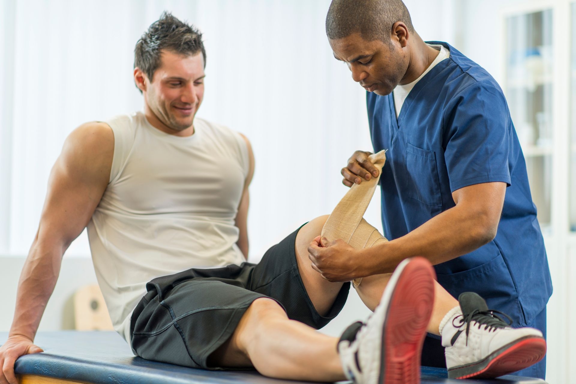
Physical examinations play a crucial role in identifying muscle weakness by assessing muscle strength, tone, and range of motion. Healthcare providers may perform strength testing, such as manual muscle testing or dynamometry, to evaluate the power of specific muscle groups. Range of motion assessments can help determine if there are any limitations in movement that may be related to muscle weakness.
When evaluating muscle weakness in patients, healthcare providers look for key symptoms and signs such as muscle fatigue, cramping, stiffness, or difficulty performing daily activities. Weakness that is progressive, asymmetric, or associated with other neurological symptoms like numbness or tingling may indicate a more serious underlying condition. Additionally, a thorough medical history, family history, and physical examination are essential in determining the cause of muscle weakness and developing an appropriate treatment plan.
Patient positioning is crucial in gluteus medius testing as it allows for accurate assessment of muscle strength and function. Proper positioning ensures that the muscle is isolated and can be tested effectively. This is important in diagnosing conditions such as gluteus medius weakness or dysfunction. Additionally, correct positioning helps in preventing compensatory movements or recruitment of other muscles during testing, which can lead to inaccurate results. By positioning the patient correctly, healthcare providers can obtain reliable data to inform treatment plans and monitor progress. Overall, patient positioning plays a significant role in the accuracy and effectiveness of gluteus medius testing.
Muscle weakness can be detected during manual muscle testing by assessing the individual's ability to generate force against resistance. The examiner applies pressure to specific muscle groups while the individual performs a specific movement, such as flexing or extending a joint. Weakness may be indicated by the individual's inability to maintain the desired position or movement against resistance. Other signs of muscle weakness during manual muscle testing include decreased muscle tone, reduced muscle bulk, and limited range of motion. Additionally, the presence of muscle atrophy, fasciculations, or tremors may also suggest underlying weakness. Overall, manual muscle testing is a valuable tool for evaluating muscle strength and identifying areas of weakness in individuals.
To test the gastrocnemius muscle in patients experiencing calf pain, a healthcare provider may perform a series of physical examinations. This may include assessing the range of motion of the ankle joint, checking for any signs of swelling or tenderness in the calf area, and evaluating the strength of the muscle through manual resistance testing. Additionally, the provider may also conduct specific orthopedic tests such as the Thompson test to assess the integrity of the Achilles tendon, which is closely related to the function of the gastrocnemius muscle. Imaging studies like ultrasound or MRI may also be utilized to further evaluate the muscle and surrounding structures. By conducting a comprehensive assessment, healthcare providers can accurately diagnose and treat conditions affecting the gastrocnemius muscle in patients with calf pain.
Finger extensor muscle strength can be measured manually using a handheld dynamometer or a pinch gauge. The examiner applies resistance against the individual's finger extension movement while they attempt to straighten their fingers against the force. The force exerted by the finger extensor muscles is then quantified in pounds or kilograms. Additionally, manual muscle testing can be performed using the Medical Research Council (MRC) scale to grade the strength of the finger extensor muscles on a scale from 0 to 5, with 0 indicating no muscle contraction and 5 indicating normal strength. This manual assessment method allows for a subjective evaluation of the individual's finger extensor muscle strength and can help in monitoring progress during rehabilitation or treatment.
Various techniques are utilized to measure finger extensor muscle strength, including hand-held dynamometers, grip strength meters, pinch gauges, and manual muscle testing. Hand-held dynamometers are commonly used to quantify the force generated by the finger extensor muscles, providing objective measurements of strength. Grip strength meters assess overall hand strength, which includes the contribution of the finger extensors. Pinch gauges specifically target the strength of the finger extensors during a pinching motion. Manual muscle testing involves a clinician applying resistance to the fingers while the individual attempts to extend them, providing a qualitative assessment of strength. These techniques collectively offer a comprehensive evaluation of finger extensor muscle strength, aiding in the diagnosis and monitoring of conditions affecting these muscles.