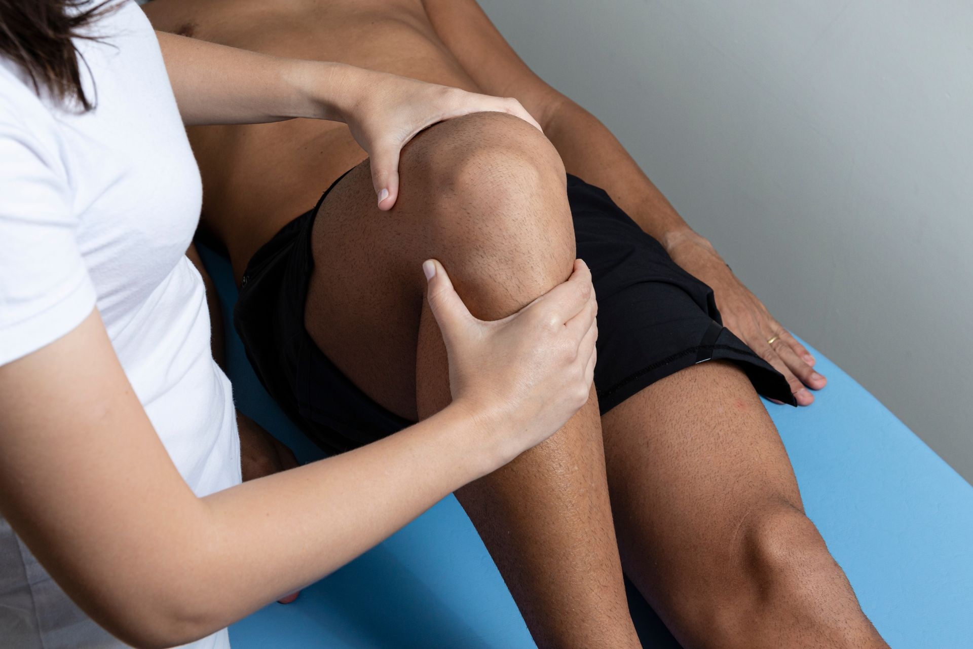

A digital goniometer measures joint angles by utilizing sensors and a digital display. The device is typically placed on the joint of interest, such as the knee or elbow, and the sensors detect the movement of the joint. The data is then processed and displayed on the digital screen, providing an accurate measurement of the joint angle. This allows healthcare professionals to assess the range of motion and track any changes over time.
There are several advantages of using a digital goniometer over a traditional goniometer. Firstly, digital goniometers provide a more precise and accurate measurement of joint angles. The digital display eliminates the need for manual interpretation, reducing the potential for human error. Additionally, digital goniometers often have the ability to store and analyze data, allowing for easier tracking of progress and comparison between different measurements. They are also more convenient to use, as they are portable and can be easily carried to different locations.
In the dynamic realm of healthcare, advancements in technology continuously reshape the landscape of physical therapy. From cutting-edge therapeutic technologies to revolutionary tools for physiotherapy, the field is witnessing a rapid evolution. In this blog, we delve into the latest trends and innovations in physical therapy, exploring advanced techniques for rehabilitation and highlighting the [...]
Posted by on 2024-02-10
In the fast-paced world we live in today, maintaining optimal health and well-being has become more critical than ever. As individuals face various challenges that limit their ability to access traditional healthcare settings, the importance of home-based solutions has risen significantly. Home exercise programs and physical therapy at home have emerged as valuable alternatives, [...]
Posted by on 2023-12-26
In the pursuit of effective pain relief and improved mobility, finding innovative treatments is crucial. Town PT in Maywood introduces Shockwave Therapy as a cutting-edge approach to addressing various musculoskeletal conditions. Discover how this revolutionary therapy could transform your journey to recovery and wellness. Understanding Shockwave Therapy Shockwave Therapy is a non-invasive treatment that [...]
Posted by on 2023-12-15
Navigating insurance coverage for physical therapy services can often feel like navigating a maze. Understanding the ins and outs of physical therapy insurance coverage, reimbursement policies, and maximizing insurance benefits is crucial for both patients and healthcare providers. In this comprehensive guide, we'll delve into the theme of physical therapy insurance coverage, comparing different [...]
Posted by on 2024-02-25
Yes, a digital goniometer can be used for both active and passive range of motion measurements. Active range of motion refers to the movement of a joint performed by the individual themselves, while passive range of motion refers to the movement of a joint performed by an external force, such as a healthcare professional. The digital goniometer can accurately measure the joint angles in both scenarios, providing valuable information about the flexibility and mobility of the joint.

While digital goniometers offer many benefits, there are some limitations and potential errors associated with their use. One limitation is that the accuracy of the measurement may be affected by factors such as the placement of the device on the joint or the positioning of the individual. It is important to ensure proper alignment and positioning to obtain accurate measurements. Additionally, digital goniometers may not be suitable for all individuals, such as those with limited mobility or certain medical conditions. It is important to consider these factors and use the device in conjunction with clinical judgment.
The accuracy of a digital goniometer generally compares favorably to other methods of measuring joint angles. Traditional goniometers rely on manual interpretation and estimation, which can introduce errors. Digital goniometers provide a more precise and objective measurement, reducing the potential for human error. However, it is important to note that the accuracy may still be influenced by factors such as the placement of the device and the positioning of the individual. Overall, digital goniometers offer a reliable and accurate method for measuring joint angles.

Yes, a digital goniometer can be used for assessing joint angles in different body parts. The device can be applied to various joints, including the knee, elbow, shoulder, and wrist, among others. This versatility allows healthcare professionals to assess the range of motion and flexibility of different joints throughout the body. By measuring joint angles in different body parts, healthcare professionals can gain a comprehensive understanding of an individual's mobility and identify any limitations or abnormalities.
Digital goniometers have a range of applications in clinical settings. They are commonly used in physical therapy and rehabilitation to assess and monitor the progress of patients recovering from injuries or surgeries. Digital goniometers can also be used in orthopedics to evaluate joint function and range of motion before and after surgical interventions. Additionally, they are utilized in sports medicine to assess athletes' joint mobility and track any changes over time. The data obtained from digital goniometers can inform treatment plans, guide interventions, and provide objective measurements for research purposes.

Telemedicine platforms have revolutionized the field of physical therapy by enabling remote diagnostic assessments with unprecedented convenience and efficiency. These platforms leverage cutting-edge technologies such as video conferencing, remote monitoring devices, and artificial intelligence algorithms to facilitate accurate and real-time assessments of patients' physical conditions. With the ability to remotely evaluate patients' range of motion, strength, and functional abilities, physical therapists can provide personalized treatment plans and interventions without the need for in-person visits. This not only saves time and travel costs for patients but also allows for continuous monitoring and adjustment of treatment plans based on real-time data. Furthermore, telemedicine platforms in physical therapy have seen advancements in the integration of wearable devices and sensors, enabling therapists to remotely track patients' progress and adherence to prescribed exercises. Overall, these advancements in telemedicine platforms have transformed the way physical therapy assessments are conducted, making it more accessible, efficient, and patient-centered.
Specialized diagnostic equipment commonly used for assessing neuromuscular junction disorders in physical therapy patients includes electromyography (EMG), nerve conduction studies (NCS), repetitive nerve stimulation (RNS), and single-fiber electromyography (SFEMG). EMG measures the electrical activity of muscles and can help identify abnormalities in the neuromuscular junction. NCS evaluates the speed and strength of nerve signals, providing information about nerve function and potential abnormalities. RNS involves delivering repetitive electrical stimuli to assess the response of muscles, helping to diagnose conditions such as myasthenia gravis. SFEMG is a highly sensitive test that evaluates the electrical activity of individual muscle fibers, aiding in the diagnosis of disorders affecting the neuromuscular junction. These specialized diagnostic tools enable physical therapists to accurately assess and diagnose neuromuscular junction disorders, guiding the development of effective treatment plans for their patients.
The specific diagnostic criteria for assessing thoracic outlet syndrome in physical therapy involve a comprehensive evaluation of the patient's symptoms, medical history, and physical examination findings. The physical therapist will look for specific signs and symptoms such as pain or discomfort in the neck, shoulder, arm, or hand, weakness or numbness in the affected limb, and changes in sensation or temperature. They will also assess for any postural abnormalities, muscle imbalances, or restricted range of motion in the neck and shoulder region. Additionally, the physical therapist may perform special tests such as the Adson's maneuver, Wright's test, or Roos test to further evaluate the presence of thoracic outlet syndrome. Imaging studies such as X-rays, MRI, or ultrasound may be ordered to rule out other potential causes of the symptoms. Overall, a thorough assessment using a combination of subjective and objective measures is essential for an accurate diagnosis of thoracic outlet syndrome in physical therapy.
Musculoskeletal ultrasound is a valuable tool in physical therapy for diagnosing tendon injuries. This imaging technique uses high-frequency sound waves to create detailed images of the musculoskeletal system, allowing physical therapists to visualize and assess the condition of tendons. By utilizing musculoskeletal ultrasound, physical therapists can accurately identify and evaluate tendon injuries such as tendinitis, tendinosis, and tendon tears. The ultrasound images provide information about the size, shape, and integrity of the tendon, as well as any abnormalities or inflammation present. This enables physical therapists to develop targeted treatment plans and monitor the progress of tendon healing. Additionally, musculoskeletal ultrasound can be used to guide therapeutic interventions such as injections or needle-based procedures, ensuring precise and effective treatment. Overall, musculoskeletal ultrasound plays a crucial role in the diagnosis and management of tendon injuries in physical therapy, enhancing the quality of care provided to patients.
Clinicians utilize functional near-infrared spectroscopy (fNIRS) as a non-invasive neuroimaging technique to diagnose cerebral impairments in physical therapy patients. By measuring changes in oxygenated and deoxygenated hemoglobin levels in the brain, fNIRS provides valuable insights into cerebral blood flow and neuronal activity. This information allows clinicians to assess the functional integrity of the brain and identify any abnormalities or impairments that may be affecting the patient's motor or cognitive functions. By analyzing the data obtained from fNIRS, clinicians can make informed decisions regarding the appropriate course of treatment and rehabilitation strategies for physical therapy patients with cerebral impairments.
The specific diagnostic protocols for identifying cervical radiculopathy in physical therapy patients involve a comprehensive assessment of the patient's medical history, physical examination, and diagnostic imaging. The physical therapist will begin by gathering information about the patient's symptoms, such as neck pain, radiating arm pain, weakness, and sensory changes. They will also inquire about any previous injuries or medical conditions that may contribute to the development of cervical radiculopathy. During the physical examination, the therapist will assess the patient's range of motion, muscle strength, reflexes, and sensation in the affected area. They may also perform special tests, such as the Spurling's test or the upper limb tension test, to further evaluate nerve root compression. Diagnostic imaging, such as X-rays, MRI, or CT scans, may be ordered to confirm the diagnosis and identify the specific location and severity of nerve compression. By utilizing these specific diagnostic protocols, physical therapists can accurately identify cervical radiculopathy in their patients and develop appropriate treatment plans.