
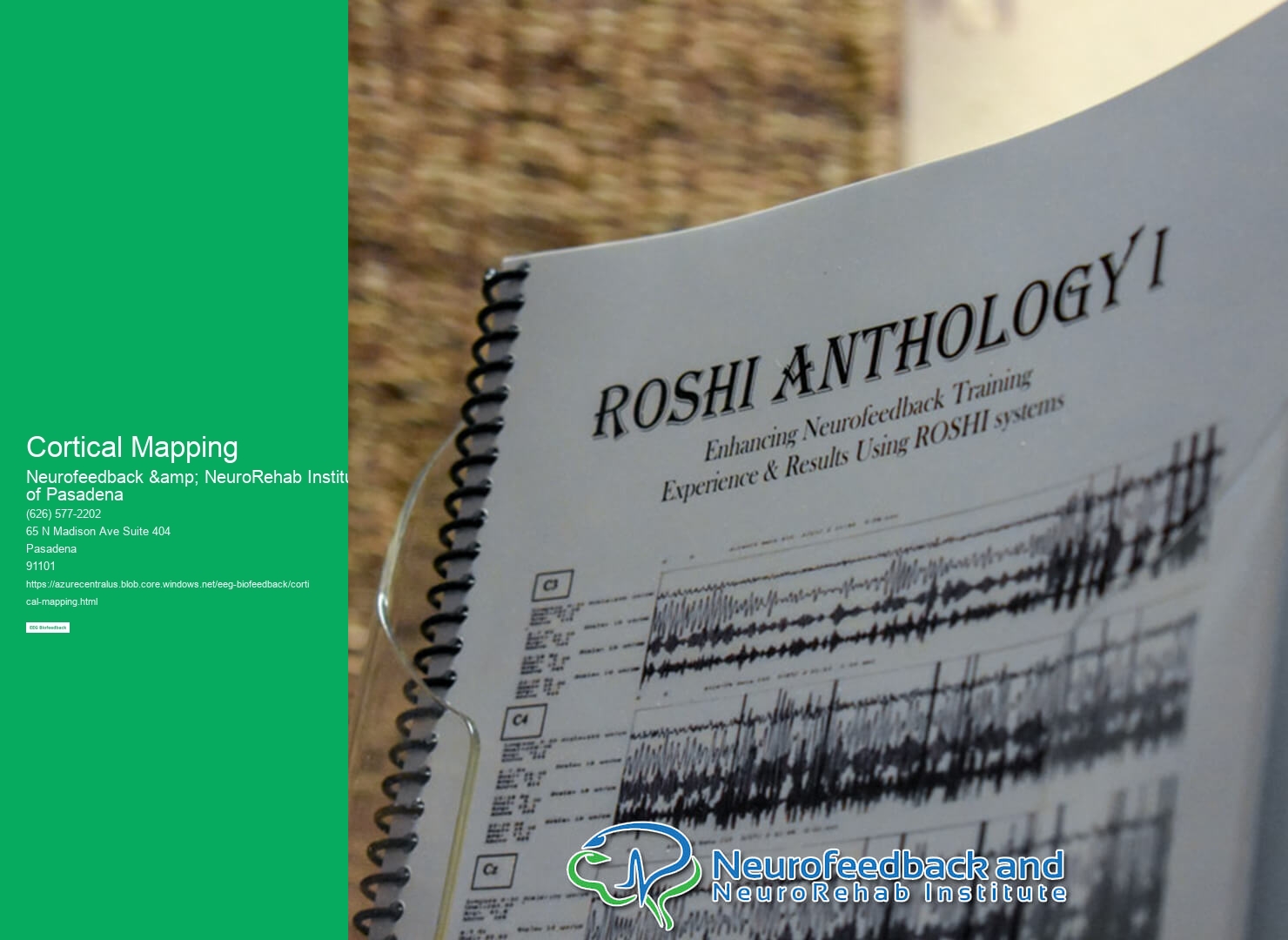
Cortical mapping is a technique used in neuroscience research to identify and understand the organization of the cerebral cortex, the outer layer of the brain responsible for higher cognitive functions. It involves mapping the different regions of the cortex and their corresponding functions. This is done by stimulating specific areas of the cortex and observing the resulting neural activity or behavioral responses. Cortical mapping can be performed using various methods, such as electrical stimulation, functional magnetic resonance imaging (fMRI), and transcranial magnetic stimulation (TMS).
There are several techniques used for cortical mapping in neuroscience research. Electrical stimulation involves applying small electrical currents to specific areas of the cortex to observe the resulting neural activity or behavioral responses. Functional magnetic resonance imaging (fMRI) measures changes in blood flow in the brain, allowing researchers to identify regions of the cortex that are active during specific tasks or functions. Transcranial magnetic stimulation (TMS) uses magnetic fields to stimulate specific areas of the cortex and observe the resulting neural activity or behavioral responses. Other techniques include optogenetics, which involves using light to control the activity of specific neurons, and single-unit recording, which involves recording the electrical activity of individual neurons.
Cortical mapping plays a crucial role in understanding brain functions and disorders. By mapping the different regions of the cortex and their corresponding functions, researchers can gain insights into how the brain processes information and controls behavior. This knowledge can help in understanding various brain functions, such as perception, language, memory, and motor control. Additionally, cortical mapping can provide valuable information about the neural basis of brain disorders, such as epilepsy, stroke, and neurodegenerative diseases. By identifying the regions of the cortex that are affected in these disorders, researchers can develop targeted treatments and interventions.
Yes, cortical mapping can be used to identify specific regions of the brain responsible for certain functions. By stimulating different areas of the cortex and observing the resulting neural activity or behavioral responses, researchers can map out the functional organization of the brain. For example, studies have shown that specific regions of the cortex are responsible for processing visual information, language, motor control, and other cognitive functions. By identifying these regions, researchers can better understand how different brain areas work together to support complex behaviors and functions.
While cortical mapping is a valuable tool in neuroscience research, it does have limitations in terms of accuracy and precision. The techniques used for cortical mapping, such as electrical stimulation and imaging methods, have inherent limitations in their ability to precisely pinpoint the exact location and function of specific cortical areas. Additionally, individual variability in brain anatomy and function can also affect the accuracy of cortical mapping. Furthermore, the interpretation of cortical mapping results requires careful analysis and consideration of various factors, such as the specific task or stimulus used, the timing of the measurements, and the potential confounding factors.

Ethical considerations are an important aspect of cortical mapping research. Invasive techniques, such as electrical stimulation and single-unit recording, involve direct manipulation of the brain and carry potential risks to the participants. Therefore, researchers must ensure that the benefits of the research outweigh the potential risks and that appropriate informed consent is obtained from the participants. Additionally, the use of animals in cortical mapping research raises ethical concerns, and researchers must adhere to strict guidelines and regulations to ensure the welfare and ethical treatment of the animals involved.
Cortical mapping contributes to the development of brain-computer interfaces and neuroprosthetics by providing insights into the neural basis of motor control and sensory processing. By mapping the regions of the cortex responsible for controlling specific movements or processing sensory information, researchers can develop algorithms and technologies that can decode and interpret neural signals. This allows individuals with motor disabilities to control external devices, such as prosthetic limbs or computer interfaces, using their brain activity. Cortical mapping also helps in understanding how the brain adapts and reorganizes after injury or disease, which is crucial for the development of effective rehabilitation strategies and interventions.
Real-time processing plays a crucial role in EEG biofeedback applications by enabling the immediate analysis and interpretation of brainwave data. This real-time analysis allows for the timely detection of specific brainwave patterns and the subsequent delivery of feedback to the user. By utilizing advanced algorithms and signal processing techniques, real-time processing can accurately identify and quantify various brainwave frequencies, such as alpha, beta, theta, and delta waves. This information is then used to provide real-time feedback to the user, helping them understand and regulate their brain activity. Additionally, real-time processing allows for the customization and adaptation of the feedback based on the user's specific needs and goals. Overall, real-time processing in EEG biofeedback applications enhances the effectiveness and efficiency of the training process, enabling users to achieve optimal brainwave patterns and improve their cognitive performance.
EEG biofeedback, also known as neurofeedback, can be administered both in-person and remotely. In-person sessions typically involve the use of specialized equipment, such as EEG sensors, which are placed on the scalp to measure brainwave activity. These sessions are usually conducted in a clinical or therapeutic setting, with a trained professional guiding the process. However, advancements in technology have made it possible to administer EEG biofeedback remotely. Remote sessions can be conducted through the use of telehealth platforms, where the client and the practitioner can connect virtually. During these sessions, the client can wear a portable EEG device that transmits the brainwave data to the practitioner in real-time. This allows for the monitoring and adjustment of the neurofeedback protocol, even when the client is not physically present in the same location as the practitioner. Remote EEG biofeedback offers convenience and accessibility, particularly for individuals who may have difficulty traveling to in-person sessions.
EEG biofeedback can indeed be integrated into virtual reality (VR) environments to enhance training. By combining the power of EEG technology with the immersive experience of VR, individuals can receive real-time feedback on their brain activity while engaging in virtual training scenarios. This integration allows for a more personalized and targeted approach to training, as the VR environment can be adjusted based on the individual's brainwave patterns. Additionally, the use of VR can enhance the engagement and motivation of the trainee, leading to improved learning outcomes. Overall, the integration of EEG biofeedback into VR environments holds great potential for enhancing training in various fields, such as sports, healthcare, and education.
EEG artifact removal techniques are seamlessly integrated into biofeedback sessions to ensure accurate and reliable data collection. These techniques involve the identification and elimination of unwanted signals or artifacts that may interfere with the interpretation of EEG signals. Common artifacts include eye blinks, muscle activity, and electrical interference. To address these artifacts, various methods are employed, such as independent component analysis (ICA), template matching, and adaptive filtering. ICA separates the EEG signals into independent components, allowing the removal of artifacts by excluding specific components. Template matching involves comparing the recorded EEG signals with pre-defined templates of known artifacts, enabling their identification and subsequent removal. Adaptive filtering techniques dynamically adjust the filter parameters to minimize the impact of artifacts on the EEG signals. By integrating these artifact removal techniques into biofeedback sessions, practitioners can obtain clean and accurate EEG data, leading to more effective and personalized biofeedback interventions.
Individuals with sleep disorders can indeed benefit from EEG biofeedback interventions. EEG biofeedback, also known as neurofeedback, is a non-invasive technique that uses real-time monitoring of brainwave activity to train individuals to self-regulate their brain function. By providing individuals with real-time feedback on their brainwave patterns, EEG biofeedback helps them learn to modify their brain activity and improve their sleep quality. This intervention has been shown to be effective in treating various sleep disorders, such as insomnia, sleep apnea, and restless leg syndrome. Through repeated sessions of EEG biofeedback, individuals can learn to achieve a more balanced and optimal brainwave pattern during sleep, leading to improved sleep quality and overall well-being.
EEG biofeedback, also known as neurofeedback, has shown promise in workplace settings for stress reduction and performance improvement. By measuring and providing feedback on brainwave activity, individuals can learn to self-regulate their brain function and achieve a state of optimal performance. This non-invasive technique has been used to address a range of workplace challenges, including stress management, attention deficits, and anxiety. Through the use of specialized equipment and training protocols, employees can learn to recognize and modify their brainwave patterns, leading to improved focus, productivity, and overall well-being. EEG biofeedback has the potential to be a valuable tool in promoting a healthy and productive work environment.