Assessing Functional Movement Patterns: A Key Component of Rehab Success

Assessing Functional Movement Patterns: A Key Component of Rehab Success

Assessing Functional Movement Patterns is essential for understanding individual capabilities and identifying areas for improvement. This process plays a key role in achieving success in rehabilitation by enhancing movement efficiency and reducing the risk of injury.

How do specific functional movement assessments influence individualized rehabilitation protocols for athletes recovering from ACL injuries?

Specific functional movement assessments play a crucial role in shaping individualized rehabilitation protocols for athletes recovering from ACL injuries by providing detailed insights into their biomechanical deficits and movement patterns. These assessments often include tests such as single-leg squat, hop tests, and dynamic balance evaluations that help identify weaknesses in strength, stability, proprioception, and coordination. By analyzing the athlete's performance during these movements, physical therapists can pinpoint areas requiring targeted intervention to restore optimal function while minimizing the risk of re-injury. For example, if an assessment reveals inadequate knee stability or poor landing mechanics during plyometric activities, tailored exercises focusing on neuromuscular training can be incorporated to enhance muscular control and joint alignment. Furthermore, understanding specific gait abnormalities through video analysis allows clinicians to design progressive loading strategies that align with each athlete’s unique recovery timeline—ensuring safe return-to-sport decisions based on evidence-based practices rather than generalized protocols. Additionally, addressing psychological components related to fear of reinjury is vital; thus incorporating mental skills training alongside physical conditioning helps build confidence in movement capabilities post-rehabilitation. Overall, leveraging data from functional movement assessments enables healthcare providers to create comprehensive rehabilitation plans centered around individual needs—ultimately facilitating effective recovery pathways for athletes striving towards pre-injury performance levels after an ACL injury.

Effective pain management strategies in rehabilitation can significantly enhance recovery outcomes and improve overall quality of life. By exploring various techniques, individuals can discover tailored approaches that suit their specific needs and promote healing. To gain deeper insights into these effective methods, visit exploring innovative approaches to recovery

In what ways can the analysis of kinetic chain dysfunctions enhance outcomes in patients undergoing post-surgical shoulder rehab?

The analysis of kinetic chain dysfunctions can significantly enhance outcomes in patients undergoing post-surgical shoulder rehabilitation by identifying and addressing imbalances, compensations, and abnormalities throughout the interconnected system of muscles, joints, and connective tissues that contribute to overall movement. By thoroughly assessing these dysfunctions, clinicians can create individualized exercise programs that target specific muscle weaknesses or tightness along the kinetic chain—such as those affecting the scapulae, thoracic spine, or even lower extremities—that may be influencing shoulder mechanics. This tailored approach helps restore proper biomechanics by promoting optimal joint alignment and enhancing neuromuscular control during functional activities like reaching overhead or lifting objects. Furthermore, understanding how proximal stability from core engagement influences distal mobility at the shoulder allows for more effective interventions aimed at improving strength while minimizing pain and risk of re-injury. Incorporating techniques such as manual therapy to improve soft tissue flexibility alongside proprioceptive training enhances coordination among musculature responsible for stabilizing the glenohumeral joint during dynamic movements. Ultimately, addressing kinetic chain issues not only facilitates a smoother recovery process but also empowers patients with strategies to prevent future injuries through education on body mechanics, posture correction exercises focusing on endurance building within key muscle groups essential for preserving shoulder health over time.

What role does assessing core stability play in identifying neuromuscular imbalances during lower extremity rehabilitation programs?

Assessing core stability is crucial in identifying neuromuscular imbalances during lower extremity rehabilitation programs because the core acts as a central hub for force transmission and balance throughout the body. When individuals have weak or dysfunctional core muscles, it can lead to compensatory movements that place excessive strain on the hips, knees, and ankles. This imbalance may result from various factors such as previous injuries, poor posture, or inadequate strength training, leading to altered biomechanics during activities like running or jumping. By evaluating an individual's ability to maintain proper alignment and control through exercises that challenge their stability—such as planks or dynamic balancing tasks—rehabilitation professionals can pinpoint specific weaknesses in muscle activation patterns across both the trunk and lower limbs. Moreover, this assessment helps determine how well these muscles engage synergistically while performing functional tasks like squatting or lunging; any discrepancies observed might indicate underlying issues such as tightness in certain muscle groups or inhibition of others due to pain signals sent from prior injuries. Consequently, addressing these neuromuscular deficits with targeted interventions not only improves overall performance but also reduces the risk of re-injury by fostering better kinetic chain integrity among all connected segments involved in movement mechanics. Thus, thorough evaluation of core stability serves not just as a diagnostic tool but also informs tailored rehabilitation strategies aimed at restoring optimal function within the entire kinetic system.

How might variations in squat mechanics inform the progression of therapeutic exercises for individuals with chronic knee pain?

Variations in squat mechanics can significantly impact the progression of therapeutic exercises for individuals suffering from chronic knee pain by providing insights into joint alignment, muscle activation patterns, and overall movement efficiency. Understanding how factors such as hip flexion, ankle dorsiflexion, and trunk positioning affect squatting techniques allows healthcare professionals to tailor rehabilitation programs that address specific deficits or compensatory strategies used by patients. For instance, a person with limited range of motion in their ankles may exhibit excessive forward lean during squats due to poor biomechanics; therefore, targeted interventions like ankle mobility drills or strength-building exercises focused on the quadriceps and hamstrings could be implemented to enhance functional capacity while minimizing discomfort. Additionally, analyzing weight distribution across the feet—whether it's more pronounced at the heels or toes—can reveal underlying issues related to stability and balance that might contribute to knee strain during activities like walking or climbing stairs. By integrating variations in squat form assessment into treatment protocols alongside modalities such as neuromuscular re-education and proprioceptive training, therapists can foster improved kinematic outcomes leading toward safer performance in daily tasks while simultaneously reducing symptoms associated with chronic knee conditions through evidence-based approaches tailored specifically for each individual's unique mechanical profile.

In which manner can the identification of compensatory movement patterns be utilized to prevent re-injury during return-to-sport phases after ankle sprains?

The identification of compensatory movement patterns plays a crucial role in preventing re-injury during the return-to-sport phases following ankle sprains. When an athlete sustains an ankle injury, they often unconsciously adjust their biomechanics to protect the injured area, leading to altered gait mechanics and muscle imbalances that can create stress on other joints like the knee or hip. By carefully assessing these compensatory movements through functional movement screenings and biomechanical analysis, coaches and rehabilitation specialists can identify specific deviations such as overpronation or reduced dorsiflexion that may predispose athletes to future injuries. Utilizing targeted interventions including strength training exercises focused on stabilizing muscles around both the ankle and lower extremities enhances proprioception and balance while correcting faulty motion patterns. Additionally, incorporating sport-specific drills helps reinforce proper biomechanics under game-like conditions ensures athletes adapt effectively without relying on maladaptive strategies. Monitoring progress with ongoing assessments allows for adjustments in rehabilitation protocols tailored to individual needs promoting optimal recovery pathways while fostering confidence when returning to competitive play; thus creating a holistic approach aimed at reducing recurrence rates of ankle sprains by addressing not only physical healing but also neuromuscular retraining essential for safe athletic participation post-injury.

Frequently Asked Questions

For effective ankle rehabilitation, it is crucial to assess specific functional movement patterns that encompass a range of biomechanical and neuromuscular components. These assessments should include the evaluation of dorsiflexion and plantarflexion capabilities during activities such as squatting or toe raises, which help determine joint mobility and muscle strength. Additionally, lateral movements like side lunges can be assessed to identify stability deficits in the peroneal muscles and overall proprioceptive balance control. Gait analysis focusing on heel strike mechanics provides insight into weight distribution and postural alignment while walking or running. Furthermore, single-leg stance tests gauge dynamic stability under various conditions, enhancing understanding of an individual's ability to maintain equilibrium during transitional movements. Incorporating these hyper-specific assessment techniques will facilitate comprehensive rehabilitation strategies aimed at restoring optimal function within the kinetic chain surrounding the ankle complex.

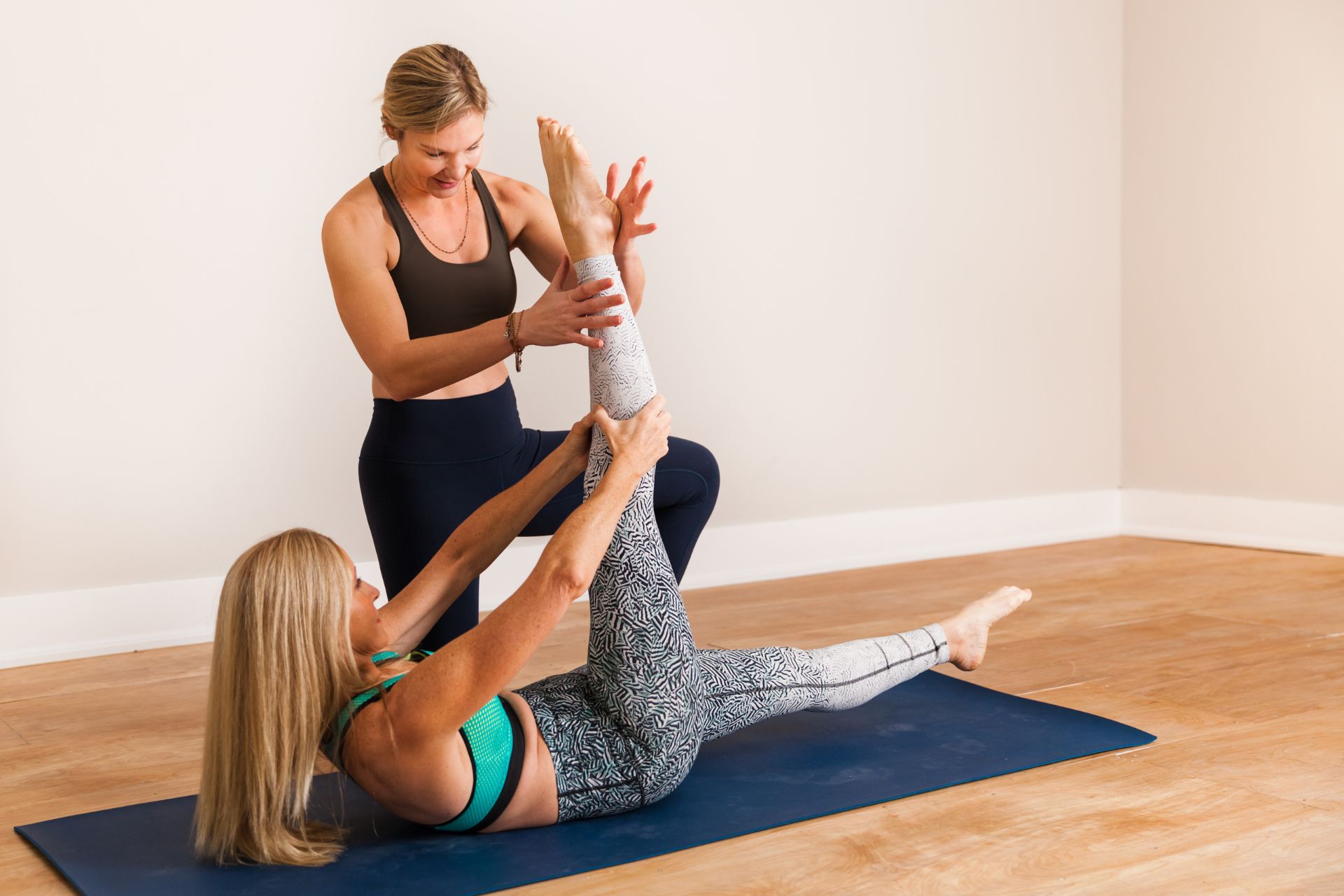
Assessing upper body mobility plays a crucial role in determining shoulder injury recovery outcomes by providing insights into the range of motion, flexibility, and stability of the glenohumeral joint. Evaluating scapular kinematics and thoracic spine mobility can highlight compensatory movement patterns that may exacerbate existing injuries or hinder rehabilitation progress. By identifying restrictions in soft tissue structures such as ligaments, tendons, and muscles surrounding the shoulder complex, healthcare professionals can develop targeted interventions to improve proprioception and neuromuscular control. Furthermore, understanding individual biomechanics allows for tailored therapeutic exercises aimed at enhancing dynamic strength while minimizing pain during functional tasks like overhead reaching or lifting. Ultimately, comprehensive assessments enable clinicians to monitor recovery trajectories more effectively and implement evidence-based strategies that promote optimal healing within musculoskeletal frameworks essential for restoring upper extremity function post-injury.

Effective tools and methods for quantifying joint stability during functional movement assessments include the utilization of inertial measurement units (IMUs), force plates, and motion capture systems that provide precise kinematic data. Electromyography (EMG) can be employed to analyze muscle activation patterns associated with stabilizing forces across joints while performing dynamic tasks such as squats or lunges. Additionally, questionnaires assessing proprioception and balance can complement these objective measures by providing insights into neuromuscular control mechanisms. The Functional Movement Screen (FMS) offers a qualitative assessment framework that identifies dysfunctional movement patterns related to joint instability. Furthermore, high-speed video analysis aids in visualizing biomechanical deviations during athletic movements, allowing practitioners to evaluate alignment and load distribution on specific articulations like the knee or ankle under varying conditions of stress. Together, these methodologies create a comprehensive approach to evaluating joint integrity within the context of functional performance capabilities.

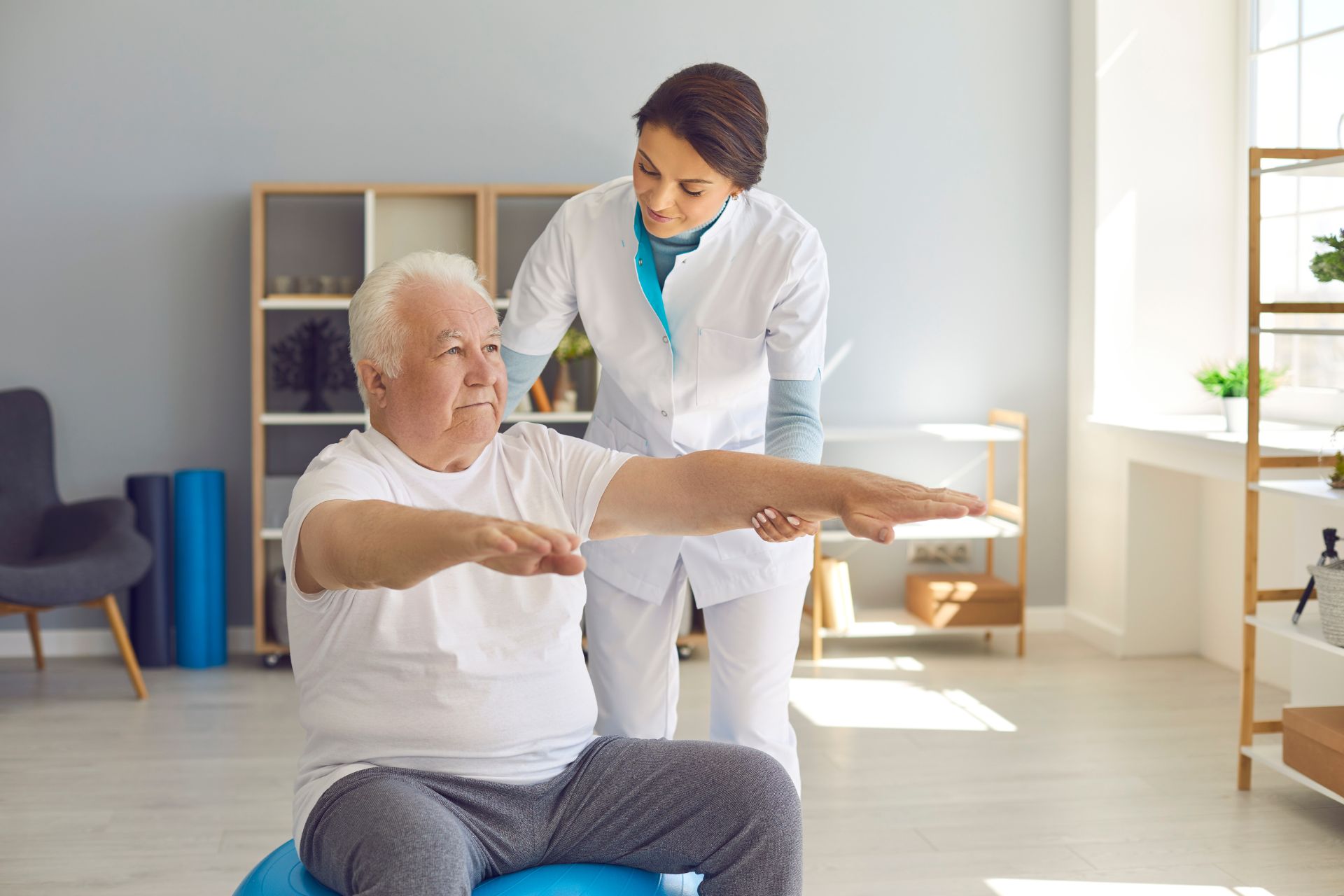
Age and fitness level significantly influence the assessment of functional movement patterns in rehabilitation programs, as they dictate an individual's musculoskeletal integrity, neuromuscular coordination, and overall biomechanical efficiency. Older adults may exhibit decreased joint flexibility, muscle atrophy, or diminished proprioception which can impede their ability to perform dynamic movements such as squats or lunges effectively. Conversely, younger individuals with higher fitness levels tend to demonstrate superior kinetic chain performance and greater muscular endurance during functional assessments like balance tests or agility drills. Tailoring rehabilitation protocols requires a nuanced understanding of how age-related physiological changes—such as reduced collagen synthesis and altered motor unit recruitment—impact movement quality while concurrently considering an individual’s baseline strength capacity and cardiovascular conditioning. This holistic approach ensures that interventions are appropriate for each demographic's unique limitations and capabilities in restoring optimal functional mobility post-injury or surgery.

Proprioception plays a critical role in the assessment of lower extremity functional movements post-injury by providing essential feedback regarding body position, movement awareness, and balance during rehabilitation. This sensory mechanism enables individuals to accurately perceive spatial orientation and joint angles, which is vital for performing complex tasks such as squatting, jumping, or cutting without compromising stability or increasing the risk of reinjury. Through proprioceptive training modalities like balance boards and closed kinetic chain exercises, clinicians can evaluate neuromuscular control and dynamic stability while assessing gait patterns and limb coordination. Moreover, quantifying proprioceptive deficits facilitates targeted interventions that enhance sensorimotor integration and functional recovery outcomes following injury events such as ACL tears or ankle sprains. By incorporating assessments focused on proprioceptive acuity within clinical protocols, healthcare professionals can better tailor rehabilitation programs to restore optimal function in patients' lower extremities efficiently.

Assessing Functional Movement Patterns: A Key Component of Rehab Success

Assessing Functional Movement Patterns: A Key Component of Rehab Success

Contact Us

Alpine Physical Therapy and Fitness

  • Address: 3130 Alpine Rd Suite 370, Portola Valley, CA 94028
  • Phone: (650) 242-0777
  • Email: alpineptfit@mail.com

© Copyright - All Rights Reserved